Baseline magnetic resonance enterography (MRE) features stratified the likelihood that Crohn’s disease (CD) strictures would persist after advanced therapy, according to a two-stage study of 187 patients and 265 MRE-defined strictures published online in Clinical Gastroenterology and Hepatology. In a temporally separated validation cohort, a summed score combining activity-related and structural imaging features had an area under the curve of 0.90 for identifying strictures that did not improve on follow-up imaging. The framework also was associated with differences in subsequent surgery-free survival.
“Most trials and real-world frameworks continue to focus primarily on active inflammation, whereas stricturing disease, a major manifestation of cumulative bowel damage, often determines clinically important outcomes, including obstruction, hospitalization, and surgery,” according to the study.
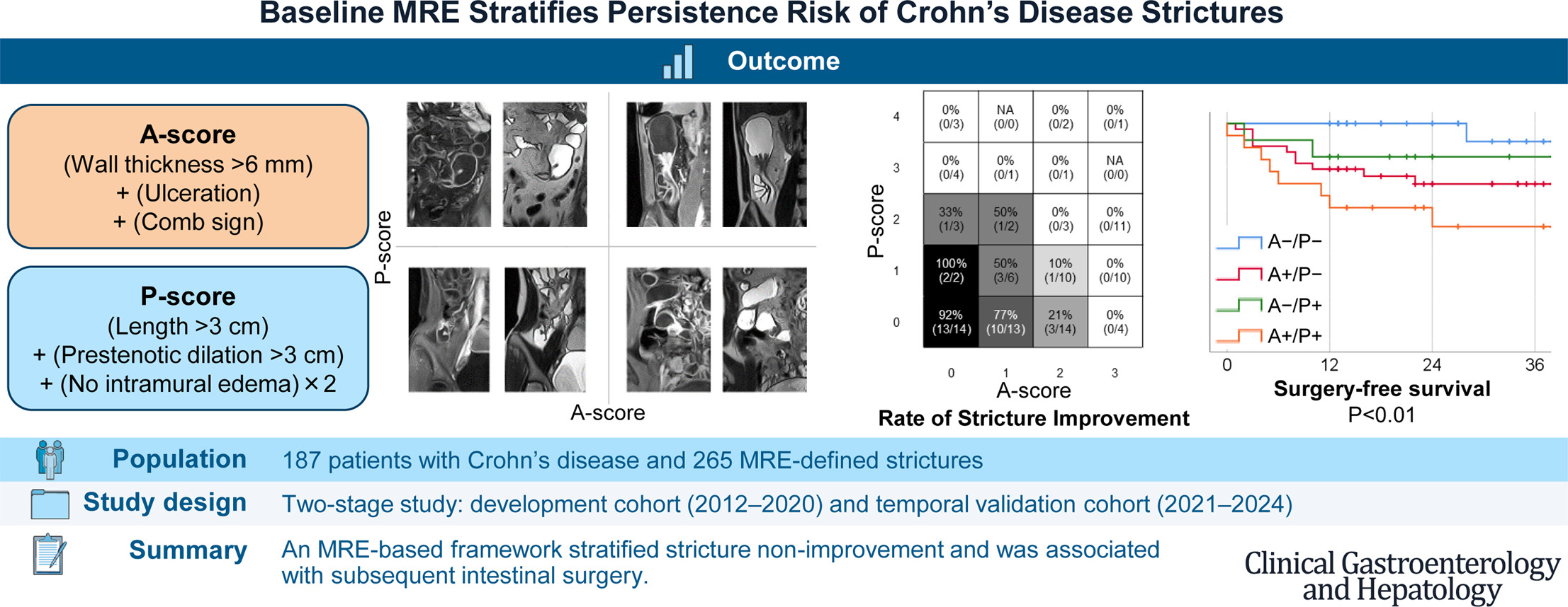
The study used a retrospective development cohort from 2012 through 2020 and a temporally separated validation cohort from 2021 through 2024, both drawn from the same prospectively maintained database at a tertiary referral hospital. Investigators developed an activity-related score and a persistence-associated structural score from baseline MRE findings, then applied the finalized scores without modification to the validation cohort. The primary endpoint was stricture-level non-improvement on follow-up MRE after initiation of advanced therapy.
The development cohort included 115 patients with 161 strictures, and the validation cohort included 72 patients with 104 strictures. Eligible patients underwent baseline MRE within two months before starting advanced therapy and follow-up MRE 12 to 24 months afterward. Patients who underwent intestinal surgery or endoscopic balloon dilation before the scheduled follow-up imaging and those whose index therapy regimen was changed before follow-up MRE, including discontinuation, switching, or dose escalation, were excluded.
Investigators defined a stricture as the simultaneous presence of bowel wall thickening greater than 3 mm, luminal narrowing, and prestenotic dilation greater than 2.5 cm. A stricture was classified as improved if it no longer met the stricture definition on follow-up imaging; otherwise, it was classified as non-improved. An abdominal radiologist and an inflammatory bowel disease physician independently reviewed the imaging while blinded to clinical information and treatment details.
At follow-up, 46 of 161 strictures (29%) in the development cohort and 34 of 104 (33%) in the validation cohort had improved. At the patient level, all baseline strictures improved in 28 of 115 patients (24%) and 16 of 72 patients (22%), respectively. Among patients with multiple strictures, responses differed among strictures within the same patient in nine of 47 (19%) in the development cohort and seven of 32 (22%) in the validation cohort.
Score development
In the development cohort, multivariable analyses identified three activity-related features associated with stricture non-improvement: bowel wall thickness greater than 6 mm, ulceration, and comb sign. These became components of the activity-related A-score. The structural P-score incorporated stricture length greater than 3 cm, prestenotic bowel diameter greater than 3 cm, and lack of intramural edema, with lack of edema weighted two points. Scores of 2 or higher were considered positive for each measure.
Validation results
In the validation cohort, the summed A+P score had an area under the curve of 0.90 for discriminating stricture non-improvement. That was significantly higher than the areas under the curve for the A-score (0.81), the P-score (0.76), the segmental Magnetic Resonance Index of Activity (MaRIA), and the segmental simplified MaRIA (0.63).
Improvement generally became less frequent as the A-score increased and remained uncommon at higher P-scores, particularly at scores of 3 or higher. Using the cutoffs locked in the development cohort, 27 of 35 strictures (77%) that were negative on both scores (A−/P−) improved. By comparison, five of 38 (13%) positive only on the A-score (A+/P−), two of 13 (15%) positive only on the P-score (A−/P+), and none of 18 positive on both (A+/P+) improved.
In the validation cohort, after adjustment for sex, disease duration, disease behavior, prior advanced therapy exposure, prior intestinal surgery, and Harvey-Bradshaw Index, a positive A-score was associated with approximately 19 times the odds of stricture non-improvement. A positive P-score was associated with approximately 20 times the odds, though the estimates were imprecise; the 95% confidence interval for the P-score ranged from 3.44 to 113.27. A-score positivity had 73% sensitivity and 85% specificity for non-improvement; P-score positivity had 41% sensitivity and 94% specificity.
Additional analyses
Exploratory analyses found no significant differences in the discriminatory performance of the summed A+P score according to stricture type, previous advanced therapy exposure, or stricture location. The area under the curve was 0.89 among native strictures and 0.93 among anastomotic strictures. It was 0.81 among strictures in patients who were advanced therapy-naive and 0.95 among those in patients previously exposed to advanced therapy. By location, the area under the curve was 0.95, 0.91, and 0.93 for colonic, terminal ileal, and proximal small-bowel strictures, respectively.
The A-score was strongly correlated with segmental MaRIA and was also correlated with segmental simplified MaRIA. In contrast, the P-score was not significantly correlated with either measure. The study authors cautioned that the findings “do not indicate that inflammation itself predicts treatment failure or that the framework directly distinguishes inflammatory from fibrotic strictures.” Instead, they described the two scores as grading complementary dimensions of baseline imaging severity.
A post hoc sensitivity analysis examined potential overlap between baseline predictors and the definition of the imaging outcome. After excluding baseline bowel wall thickness and prestenotic bowel diameter, multivariable analysis in the development cohort retained stricture length, lack of intramural edema, ulceration, comb sign, and perienteric fat stranding. A reduced non-overlapping score constructed from these five features remained associated with stricture non-improvement in the validation cohort and showed an area under the curve of 0.81. The drop in discrimination indicated that “part of the original score’s performance reflected baseline imaging severity and proximity to the follow-up thresholds,” according to the study. However, the findings “were not explained solely by predictor-outcome overlap.”
Surgical outcomes
In a secondary analysis, stricture status on follow-up imaging was associated with subsequent intestinal surgery, defined as bowel resection or strictureplasty. In the development cohort, surgery occurred in 34% of non-improved strictures compared with 2% of improved strictures during a median follow-up of 58 months. In the validation cohort, corresponding rates were 29% and 3% during a median follow-up of 22 months. Surgery-free survival in the validation cohort also differed across the four baseline A/P groups, with the A−/P− group having the lowest observed risk and the A+/P+ group the highest.
Clinical implications
None of the final score components “required contrast-enhanced or diffusion-weighted imaging, which may facilitate routine implementation,” according to the study. “Baseline magnetic resonance enterography may help identify strictures requiring closer monitoring, although prospective external validation is needed before the framework can guide treatment decisions in practice,” the study authors wrote.
“The framework identified strictures with different risks of persistence and subsequent surgery but should be interpreted as a baseline imaging severity and risk-stratification tool rather than as a treatment-response or inflammation-fibrosis classifier,” the study authors concluded. “If externally validated, this approach may support more individualized monitoring and management of CD strictures.”
Study limitations
The authors noted several limitations. The validation cohort was modest in size, and broader generalizability requires confirmation in larger and more diverse populations. Reproducibility may also depend on experienced imaging interpretation in specialized settings. The score composition, weighting, and cutoffs were partly data-driven, and comparisons with existing MRE activity indices were post hoc. Two score components, bowel wall thickness and prestenotic bowel diameter, overlapped conceptually with the follow-up stricture definition, and the reduced score that excluded them was derived post hoc. The primary outcome was binary and did not capture partial improvement or mild residual narrowing. The P-score was not validated against histologic fibrosis. In addition, excluding patients who underwent surgery or treatment modification before follow-up imaging may have introduced selection bias by removing some patients at highest risk.
The authors reported no conflicts of interest and no study funding.