A new AGA clinical practice update, published online in Clinical Gastroenterology and Hepatology, outlines when ampullary adenomas may be suitable for endoscopic papillectomy and when features such as more than 1 cm of ductal extension or suspected cancer call for surgical or multidisciplinary review.
According to the update, 20% to 40% of ampullary adenomas harbor cancer.
The expert review includes 15 best practice statements on diagnosis, treatment, and follow-up. First author Monique Barakat, MD, PhD, a pediatric and adult gastroenterologist at Stanford University School of Medicine, and colleagues based the advice on published research and expert opinion. Because they did not systematically review the evidence, they did not formally rate its quality or how strongly they supported the considerations.
“While ampullary neoplasms are relatively rare (accounting for 0.6% to 0.8% of GI cancers), their incidence among young adults has risen over the past 20 years,” Dr. Barakat told GI & Hepatology News. “Despite this trend and the fact that five-year survival rates range dramatically from 20% to 75% based on stage at diagnosis, there has historically been a relative lack of standardized guidance on best practices for managing these lesions compared to other GI neoplasms.”
Who is a candidate for papillectomy?
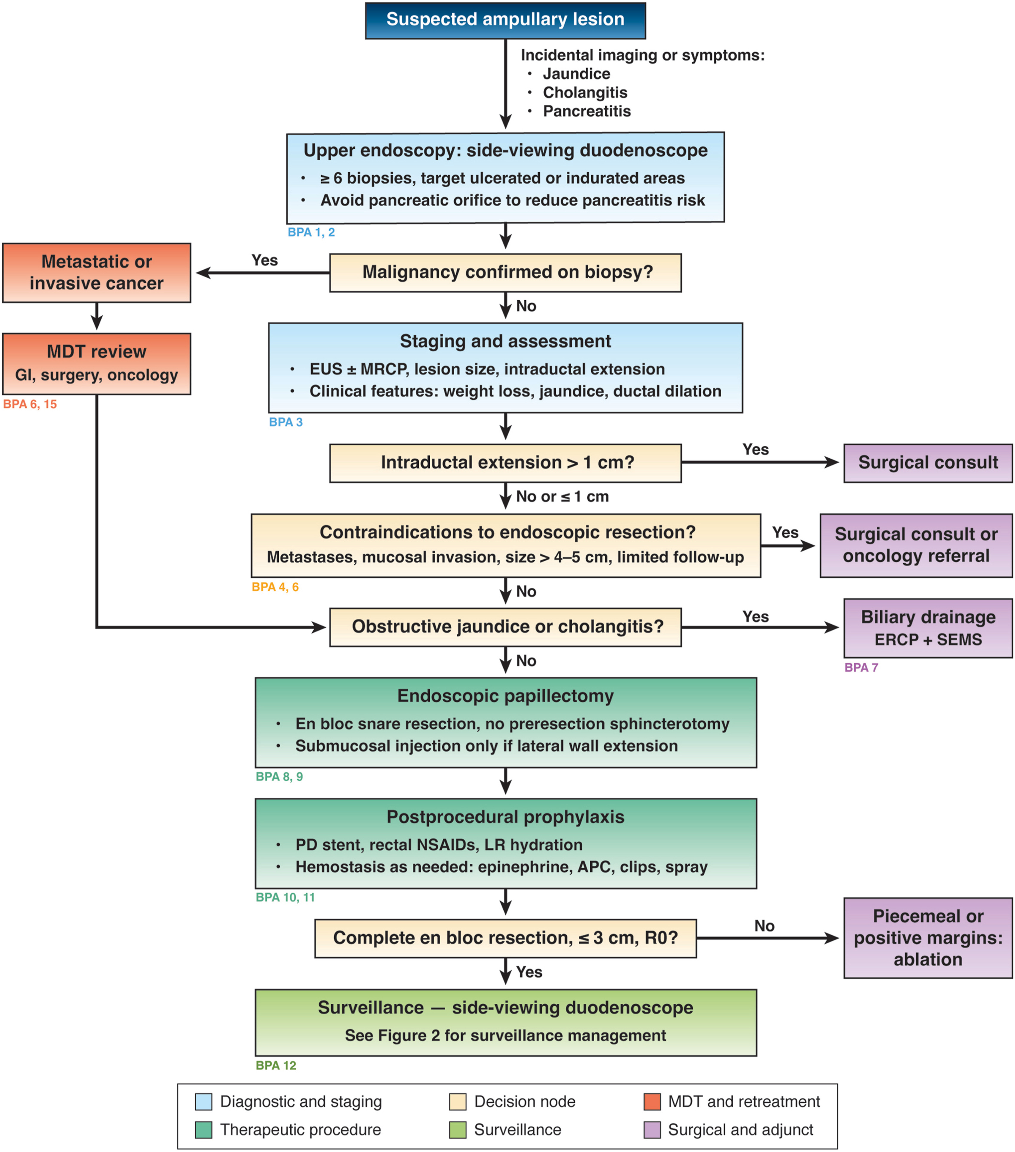
The authors recommend using a side-viewing duodenoscope to examine suspected ampullary adenomas because it provides a better view of the ampulla and better access for biopsy than a standard gastroscope.
Given the risk of cancer, they advise taking at least six biopsies, particularly from areas that are ulcerated or feel firm. They also advise avoiding the pancreatic duct opening during biopsy to reduce the risk of pancreatitis.
The authors advise considering endoscopic ultrasound for adenomas that may be removed endoscopically, to stage the lesion and determine whether it extends into the bile or pancreatic duct, except for lesions smaller than 1 cm with no worrisome features. Magnetic resonance cholangiopancreatography can be used as an adjunct to assess duct involvement, according to the update.
Several features can help determine whether endoscopic papillectomy is appropriate. According to the update, those that suggest an adenoma is benign and most amenable to papillectomy include well-defined margins, size less than 4 to 5 cm, tissue that does not bleed easily, softness when probed with the endoscope, and extension of 1 cm or less into a duct.
For adenomas extending more than 1 cm into a duct, the authors suggest consultation for possible surgical resection. They advise against papillectomy when cancer has spread, invasive cancer extends beyond the mucosa, or duct involvement exceeds 1 cm in a patient who can undergo surgery.
Even when biopsies do not show cancer, certain features should raise concern before papillectomy, the authors write. These include notable weight loss; jaundice; lesions larger than 4 cm that are friable, ulcerated, or fixed; and substantial ductal dilation with an abrupt cutoff at the ampulla on imaging.
Because robust evidence-based criteria for selecting patients for papillectomy have not been established, the decision is typically individualized based on lesion characteristics, the patient’s clinical status and preferences, and available endoscopic expertise, according to the update.
Preventing complications and recurrence
When papillectomy is appropriate, the authors advise removing the lesion in one piece with a snare when possible. They also advise against biliary and pancreatic sphincterotomy before papillectomy because electrocautery artifact and bleeding can hinder complete tissue assessment. Submucosal injection is generally not required, according to the update, but may help when the lesion spreads laterally along the duodenal wall.
To reduce the risk of pancreatitis after papillectomy, the update advises prophylactic pancreatic duct stenting, rectal nonsteroidal anti-inflammatory drugs, and high-volume hydration with lactated Ringer’s solution, adjusted for the patient’s cardiac status and size.
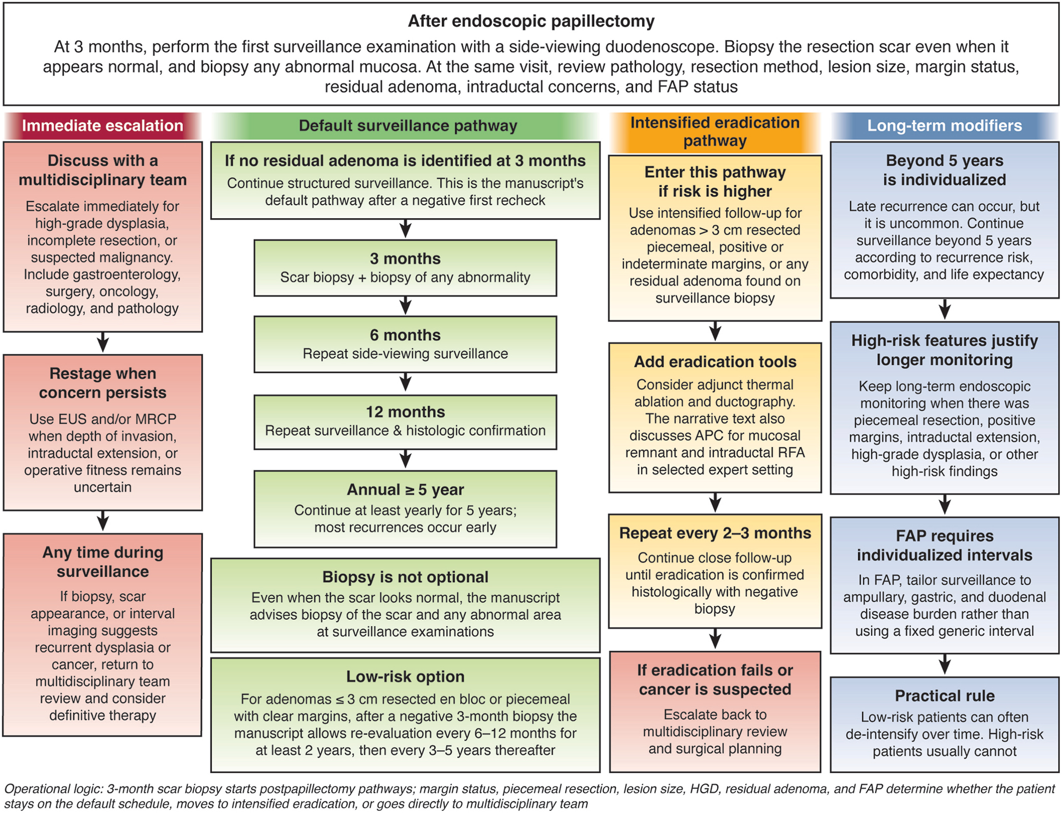
The authors advise beginning surveillance three months after papillectomy with side-viewing duodenoscopy and biopsy of the resection scar — even when it appears normal — and any abnormal areas. If no residual adenoma is found, they recommend examinations at six and 12 months, then annually for at least five years. Beyond five years, the update advises individualizing surveillance based on recurrence risk, other health conditions, and life expectancy.
For adenomas larger than 3 cm removed piecemeal or with positive margins, the authors advise considering adjunct thermal ablation and ductography, with follow-up every two to three months until biopsy confirms eradication. According to observational data and a 2024 meta-analysis cited in the update, intraductal radiofrequency ablation had a short-term clinical success rate of about 76% and a biliary stricture rate of about 22%.
Patients with familial adenomatous polyposis have higher adverse event and recurrence rates after papillectomy, and their ampullary adenomas progress less often than those in other patients, at a rate of 11% to 14%, according to the update. For that reason, some experts recommend surveillance with biopsies instead of removal when there is no rapid growth or high-grade dysplasia, though the authors note this risks missing unsampled high-grade dysplasia or cancer. The update advises tailoring surveillance intervals to disease burden in the ampulla, duodenum, and stomach.
For patients with high-grade dysplasia, incomplete removal, or suspected cancer, the authors recommend involving “a multidisciplinary team that includes gastroenterologists, surgeons, and oncologists.”
“A major message of this update is the importance of quality and safety across the entire treatment continuum,” Dr. Barakat said. “Increasing incidental detection due to broader use of cross-sectional imaging and upper endoscopy makes it crucial to establish clear, standardized protocols now to optimize early detection, endoscopic resection, and postprocedural care.”
The maximum lesion size for endoscopic resection remains controversial, according to the authors. They also note that rectal anti-inflammatory drugs have not been studied specifically in papillectomy, most studies of treatment for bleeding after papillectomy are retrospective, and reports of recurrence beyond five years are limited.
Dr. Barakat reported research support from the Cystic Fibrosis Foundation, coauthors reported consulting for or receiving personal income from Boston Scientific and Gore, and Dr. Barakat and one coauthor serve as editors of Gastrointestinal Endoscopy.

Expert insight
Dr. Barakat highlighted key takeaways from the clinical practice update for GI & Hepatology News.
Which lesion features should lead physicians to choose surgery rather than endoscopic papillectomy?
Dr. Barakat: Features that favor surgical consultation or resection over endoscopic papillectomy include relative contraindications and concerning features: lesions larger than 4 cm in size, friable or fixed lesions, early-stage/T1 cancers, notable clinical signs like significant weight loss or jaundice, and substantial ductal dilation with an abrupt cutoff.
When can endoscopic ultrasound (EUS) be safely omitted during the evaluation of an ampullary adenoma?
Dr. Barakat: EUS assessment can be safely omitted for lesions smaller than 1 cm that present with no worrisome clinical or endoscopic features (such as friability, ulceration, or ductal dilation).
Which patients need surveillance beyond five years after endoscopic resection, and how should follow-up be individualized?
Dr. Barakat: Routine annual surveillance beyond five years is not necessary for average-risk patients who have achieved durable, negative surveillance examinations. However, long-term monitoring beyond five years should be continued, or considered, for patients with high-risk features, including initial piecemeal resection, positive or indeterminate resection margins, intraductal extension, high-grade dysplasia, or familial adenomatous polyposis.