An individualized exclusion diet based on intestinal changes observed with confocal laser endomicroscopy (CLE) did not significantly improve symptoms more than a sham diet in patients with irritable bowel syndrome (IBS), a randomized crossover trial found.
Among 31 patients in the primary analysis, 41.9% responded to the targeted diet, compared with 35.5% during the sham diet (odds ratio, 1.33; 95% CI, 0.46-3.84; P = .60). The trial was stopped early after the treatment effect met a prespecified criterion for futility.
The findings, reported in Gastroenterology, challenge the use of CLE to select dietary triggers in IBS, the authors wrote. CLE provides real-time microscopic images of the intestinal lining during endoscopy. Earlier uncontrolled studies had reported symptom improvement when patients avoided foods associated with acute mucosal alterations.
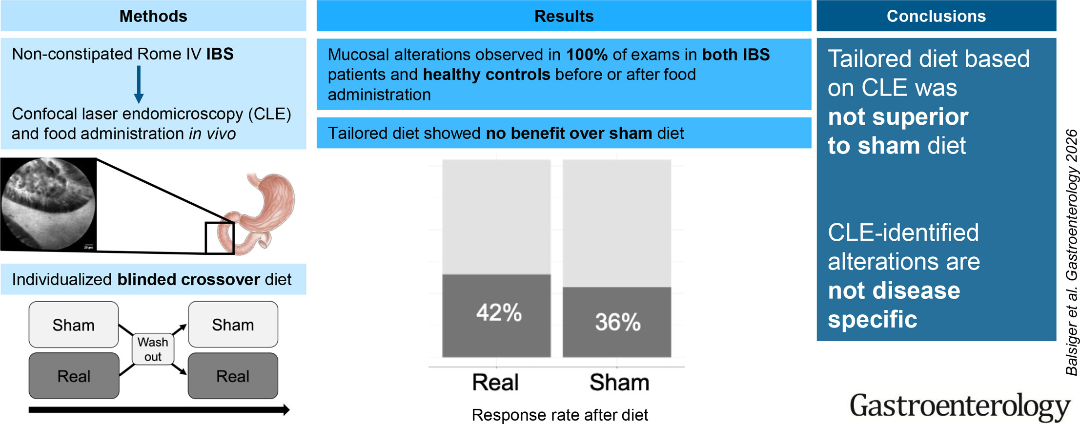
For the double-blind, controlled crossover study, first author Lukas Michaja Balsiger, PhD, of the TARGID Translational Research Group for Gastrointestinal Disorders at KU Leuven, Belgium, and colleagues enrolled 33 patients with nonconstipation-predominant IBS at University Hospitals Leuven between October 2021 and March 2024.
All met Rome IV criteria and had an IBS Symptom Severity Score (IBS-SSS) of at least 175. The median age was 33 years, 85% were women, and the median IBS-SSS was 320. Fifty-two percent also had functional dyspepsia. Patients with organic gastrointestinal disease or immunoglobulin E sensitization to any of the tested foods were excluded.
During CLE, investigators sequentially applied solutions containing wheat, milk, fish or seafood, nuts, soy, and egg white directly to the duodenal lining in random order. The physician assessing the microscopic images was blinded to which food had been administered.
The mucosa was classified as altered when more than two of four examined sites showed both fluorescein leaking into the intestinal lumen and the appearance of luminal particles. Testing stopped when an alteration was observed. Patients in whom an alteration occurred before at least three foods had been administered could undergo a second CLE examination, but no participant underwent more than two examinations. As a result, not every food was tested in every patient.
Patients then followed two four-week exclusion diets separated by a two-week washout period. During the targeted diet, they avoided up to two foods associated with CLE-detected alterations. During the sham diet, they avoided up to two foods that had not produced alterations.
If no nonreactive food was available for use in the sham diet, investigators randomly selected one or two untested foods. Neither the patients nor the investigator providing dietary instructions knew whether each diet was targeted or sham.
The primary outcome was a decrease of at least 50 points on the IBS-SSS after four weeks. Among the 31 patients who completed both diet periods, 13 responded during the targeted diet and 11 during the sham diet. The investigators found no statistically significant effect of treatment order and no evidence of carryover from the first diet period to the second.
Median IBS-SSS declined by 30 points during the targeted diet and 20 points during the sham diet, also a nonsignificant difference (P = .70).
A sensitivity analysis included 26 patients with complete data who had received a true sham diet rather than one based on untested foods. Half responded during the targeted diet, compared with 42.3% during the sham diet (odds ratio, 1.36; 95% CI, 0.46-4.07; P = .58). CLE-detected alterations had a sensitivity and specificity of 54% for predicting response to food exclusion in this analysis.
Among patients in the sensitivity analysis, the response rate to the targeted diet was 58% in those with functional dyspepsia and 43% in those without it, a difference that was not statistically significant. No food-specific comparison showed a statistically significant difference in response between targeted and sham exclusion, although the number of patients in individual food groups was small.
Daily symptom diaries also showed no significant differences between targeted and sham diets in changes in abdominal pain duration or severity, bloating, flatulence, or days with fecal urgency. Self-reported adherence was high and similar between the two diet periods.
CLE-detected mucosal alterations occurred during all 54 examinations performed among the 33 patients. They appeared before any food was administered in six examinations, or 11%, and after food administration in the remainder. Alterations occurred in 50% of examinations in which milk was administered, 43% involving fish, and 35% involving egg white.
Investigators subsequently performed the same CLE procedure in 15 age- and sex-matched healthy volunteers without gastrointestinal symptoms or food-specific immunoglobulin E sensitization. Mucosal alterations occurred in all 15 examinations, including 13% in which they appeared before any food was administered. The authors reported that the pattern was similar to that seen in patients with IBS.
The investigators also found no significant changes from baseline in ex vivo epithelial permeability or in measured markers of local mast-cell or eosinophil activation after the CLE-detected alterations. The authors wrote that the alterations may represent nonspecific mucosal responses to food exposure or mechanical stimulation rather than pathologic reactions.
The trial was stopped early after a prespecified futility analysis. The protocol defined futility as an odds ratio of 1.5 or less for response with the targeted versus sham diet; the observed odds ratio was 1.33. Early termination left the study with a relatively small sample and lower statistical power than if enrollment had continued to its originally planned target.
Other limitations included incomplete testing of all foods in each patient, administration of unprocessed foods directly to the duodenum rather than exposure after normal cooking and gastric digestion, reliance on self-reported dietary adherence, and the four-week duration of each diet. Patients knew which foods they were avoiding, although they did not know whether they were following the targeted or sham diet. The authors also noted that the sham exclusion diet was not a truly inert placebo.
KU Leuven internally funded the study through a Methusalem grant to Jan Tack. Individual investigators reported support from the Swiss National Science Foundation, the Novartis Foundation for Medical-Biological Research, and the Research Foundation Flanders. The authors declared no conflicts of interest.

Expert Insight
GI & Hepatology News spoke with Dr. Balsiger about the study.
How should these findings change the use of CLE to identify dietary triggers in patients with IBS?
Dr. Balsiger: Based on this first randomized controlled trial, there is no evidence that this technique helps identify dietary triggers.
Whether patients excluded foods that caused visible reactions or foods that did not cause reactions - the dietary intervention provided the same amount of symptom improvement.
These findings suggest that this approach is not useful to identify dietary triggers in clinical practice using the current approach.
Why did excluding foods that caused mucosal changes produce no greater symptom improvement than the sham diet?
Dr. Balsiger: We are not sure why this is the case. However, we found that mucosal changes after food administration occurred in healthy controls as well.
Finding the same patterns in healthy, asymptomatic controls indicates that these changes are indeed a physiological reaction to food rather than a pathological reaction.
The mucosal changes we were looking for were the same changes that other investigators have been looking at and we have previously validated: leaking of contrast agent and appearance of particles. It remains to be seen whether other changes might be linked with symptom generation.
Do the study’s small sample size and incomplete testing of all foods leave open a role for this approach in selected patients?
Dr. Balsiger: Given that healthy volunteers also showed changes strongly suggests a physiological normal reaction to food. Therefore, we currently see no role for the current approach in routine clinical practice. As mentioned, other identifiable changes might be linked with symptom generation in the future, but this requires further research which should be carried out within prospective study protocols.
Is there anything else you'd like to say about this work?
Dr. Balsiger: The perspective of performing microscopy in real-time in a living human being is a very exciting technique that opens a huge avenue of research. Before using it in clinical practice, it will be important to understand what changes we actually see and what the mechanisms underlying these changes really are.